
Reframing endometriosis
Endometriosis has traditionally been described as a chronic gynaecological condition characterised by the presence of endometrial-like tissue outside the uterus.¹ Its most recognised symptoms include dysmenorrhoea, chronic pelvic pain, non-menstrual pelvic pain, dyspareunia and infertility.¹–³ Yet this traditional definition increasingly feels too narrow. It describes where lesions may be found, but not the full biological, clinical or human reality of the disease.
Endometriosis affects an estimated 176 million women worldwide, and diagnosis still takes an average of around eight to ten years from first medical presentation.⁴ During this period, many women move between general practice, gynaecology, gastroenterology, fertility services, pain clinics and emergency care without a clear explanation for their symptoms. This delay is not simply a diagnostic inconvenience. It has consequences for fertility, education, employment, mental health, relationships, healthcare utilisation and quality of life.
For decades, the dominant explanation for endometriosis has been Sampson’s theory of retrograde menstruation, first proposed in 1927. This theory suggests that menstrual tissue flows backward through the fallopian tubes into the pelvic cavity, where it implants and grows.³ It remains an important component of the disease model, but it cannot explain everything. Many women experience retrograde menstruation without developing endometriosis. Endometrial-like tissue has also been identified outside the pelvis, including in distant anatomical sites, raising the possibility of lymphatic, vascular, neural or systemic mechanisms of dissemination.⁵
The clinical picture is equally complex. Some women with visible disease are relatively asymptomatic, while others experience disabling pain, gastrointestinal symptoms, fatigue, brain fog and systemic manifestations that appear disproportionate to lesion burden. This disconnect between lesion location and symptom severity is one of the reasons endometriosis should no longer be viewed only as a local pelvic disorder.
The disease is now better understood as a chronic, inflammatory, hormonally influenced and potentially systemic condition. Immune dysfunction, inflammatory signalling, genetic susceptibility, altered pain processing and neuro-gastrointestinal involvement may all contribute to its presentation. This raises a fundamental question: is endometriosis truly just a gynaecological disorder?
The association between endometriosis and autoimmune disease
The association between endometriosis and autoimmune disease has been proposed in several studies, including work by Kvaskoff et al., Shigesi et al. and Aziz et al.³,⁶,⁷ Current research recognises that endometriosis has a strong heritable component, prompting investigation into whether genes associated with immune-mediated and autoimmune conditions may also contribute to endometriosis risk.⁸
A recent study by Shigesi et al. expands this question by examining both clinical and genetic associations between endometriosis and immune-mediated or inflammatory disorders, including osteoarthritis, rheumatoid arthritis, multiple sclerosis, coeliac disease and psoriasis.⁹ The study suggests that women with endometriosis may have a 30–80% increased risk of developing certain autoimmune or immune-mediated diseases.⁹ This does not prove that endometriosis is itself an autoimmune disease, but it strengthens the argument that immune biology is central to the condition.
Earlier research had identified possible overlap between genetic markers associated with rheumatoid arthritis and endometriosis, but such studies were limited by sample size and population diversity.⁸ The newer Shigesi dataset, drawing on thousands of endometriosis cases and tens of thousands of immunological disease cases, gives greater weight to the possibility of shared biological pathways.⁹
This matters because classification shapes care. If endometriosis is seen only as a reproductive disorder, then the clinical response is likely to remain centred on pelvic imaging, hormonal suppression, fertility management and surgery. If it is recognised as a systemic inflammatory and immune-associated disease, then the clinical lens broadens. It becomes reasonable to ask whether women with endometriosis should be monitored for immune-mediated comorbidities, whether biomarkers could identify disease subtypes, and whether treatments should extend beyond conventional hormonal and surgical pathways.
A neuro-gastrointestinal dimension: why EndoSure is relevant
One of the most interesting developments in this reframing is the emerging work around gastrointestinal myoelectrical activity, or GIMA, as a biomarker of endometriosis.
Endometriosis is frequently associated with gastrointestinal symptoms, including bloating, nausea, abdominal pain, altered bowel habits, early satiety and symptoms that are often mislabelled as irritable bowel syndrome. In 1998, Mathias and colleagues reported that women with laparoscopically confirmed endometriosis and gastrointestinal symptoms demonstrated a characteristic neuromuscular abnormality of the gastrointestinal tract, described as ampulla of Vater–duodenal wall spasm, a “seizure equivalent” of the enteric nervous system.¹⁰ This was an important conceptual shift. It suggested that endometriosis may alter hollow-organ function through biochemical, inflammatory or neuroenteric mechanisms, rather than producing symptoms only through local pelvic lesions.
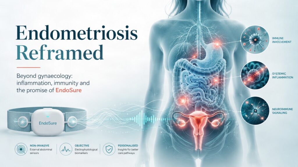
EndoSure builds directly on this concept. The technology uses non-invasive electroviscerography, recorded via abdominal electrodes, to detect abnormal gastrointestinal myoelectrical activity associated with endometriosis. The proposed biological mechanism is that endometriotic lesions secrete prostaglandin E2 and prostaglandin F2α, which may induce distinctive small-bowel smooth muscle activity and abnormal frequency patterns detectable as a GIMA biomarker.¹¹,¹²
This is important because it reframes the diagnostic question. Instead of asking only, “Can we see the lesion?” EndoSure asks, “Can we detect a physiological signature of the disease?” That distinction is crucial. Many current diagnostic approaches are better at identifying advanced or deep disease than superficial or early disease. MRI and transvaginal ultrasound can be highly useful in expert hands, particularly for deep endometriosis, but they are less reliable for superficial peritoneal disease. A physiological biomarker could therefore occupy a different place in the pathway: not as a replacement for clinical judgement, imaging or surgery, but as a potential triage and decision-support tool.
The 2024 GIMA biomarker study by Noar, Mathias and Kolatkar reported that electroviscerography with AI-derived threshold scoring distinguished participants with and without endometriosis, with high reported sensitivity, specificity, positive predictive value and negative predictive value.¹¹,¹² Additional preliminary clinical evaluations have explored EndoSure in gynaecology consultation settings and specialised endometriosis centres. Some early datasets suggest ease of use and high sensitivity, although specificity has varied between cohorts, emphasising the need for larger, independently validated, multicentre studies before the test can be positioned as a definitive diagnostic standard.¹³–¹⁵
Even with this caution, the conceptual value is significant. EndoSure is not merely another diagnostic device. It reflects a broader biological idea: that endometriosis may generate measurable physiological disturbances beyond the pelvis. If validated, such a test could help move endometriosis from a late-stage surgical diagnosis towards an earlier, pathway-based, non-invasive diagnostic model.
Why this matters clinically
The current diagnostic pathway remains too slow, too invasive and too dependent on specialist access. Many women experience symptoms for years before receiving a diagnosis. Laparoscopy remains important, but it is invasive, costly and not always immediately available. Imaging is valuable, but its performance depends heavily on disease phenotype, operator expertise and anatomical location.
A non-invasive test such as EndoSure could be clinically important if it helps identify patients who should be fast-tracked to specialist endometriosis care, advanced imaging, laparoscopy, fertility counselling or multidisciplinary management. It could also help reduce repeated consultations, inappropriate reassurance, unnecessary investigations and the misclassification of symptoms as purely functional gastrointestinal disease.
The broader opportunity is not simply earlier diagnosis. It is earlier classification. A woman with pelvic pain, bowel symptoms, fatigue and fertility concerns should not have to wait years for someone to connect the pattern. A validated biomarker could help clinicians recognise endometriosis as a multisystem disease signal, rather than a diagnosis of exclusion reached only after prolonged suffering.
Should we reframe endometriosis?
The answer is yes but carefully.
Endometriosis should not be simplistically rebranded as an autoimmune disease without stronger causal evidence. However, the growing literature on immune associations, genetic overlap, chronic inflammation and systemic symptoms makes it increasingly difficult to defend the idea that endometriosis is only a gynaecological disorder.
A more accurate framing may be:
Endometriosis is a chronic, inflammatory, hormonally responsive, neuro-gastrointestinal and immune-associated disease with reproductive, pelvic, systemic and quality-of-life consequences.
This broader framing creates new opportunities for research and care. Genetic and immune profiling could help identify subtypes of disease. Biomarkers may help predict disease severity, recurrence, treatment response or comorbidity risk. Technologies such as EndoSure may help identify physiological signatures that are not visible on standard imaging. Patient pathways could become multidisciplinary, involving gynaecologists, gastroenterologists, pain specialists, immunologists, fertility specialists, psychologists and primary care physicians.
It also has policy implications. If endometriosis is framed only as “period pain”, it will continue to be underfunded, underdiagnosed and undertreated. If it is recognised as a chronic systemic disease affecting productivity, fertility, mental health and long-term health outcomes, then it becomes a public health issue, a women’s health equity issue and a health economics issue.
Perhaps most importantly, this reframing validates lived experience. Symptoms such as fatigue, brain fog, gastrointestinal dysfunction, joint pain and widespread pain are often dismissed because they do not fit neatly into a narrow gynaecological model. A systemic disease framework gives women the language and legitimacy to describe what they are experiencing. It also gives clinicians a more coherent framework for listening.
Endometriosis is not just “bad period pain”. It is not just a fertility condition. It is not just a surgical disease. It is a complex chronic disorder that deserves the same diagnostic urgency, research investment and pathway innovation as other inflammatory and immune-mediated diseases.
In the next article, Reframing endometriosis — Part 2, we will explore how immune cell dysfunction, prostaglandin signalling, hormonal-immune interactions and gastrointestinal myoelectrical activity may help explain the multisystem nature of endometriosis — and how this understanding could reshape public health policy, diagnostic pathways and long-term disease management.
References
- Farquhar, C. M. (2007). Endometriosis. BMJ, 334(7587), 249–253. https://doi.org/10.1136/bmj.39073.736829.be
- Ballard, K. D., Seaman, H. E., de Vries, C. S., & Wright, J. T. (2008). Can symptomatology help in the diagnosis of endometriosis? Findings from a national case–control study—Part 1. BJOG: An International Journal of Obstetrics and Gynaecology, 115(11), 1382–1391. https://doi.org/10.1111/j.1471-0528.2008.01878.x
- Shigesi, N., Kvaskoff, M., Kirtley, S., Feng, Q., Fang, H., Knight, J. C., Missmer, S. A., Rahmioglu, N., Zondervan, K. T., & Becker, C. M. (2019). The association between endometriosis and autoimmune diseases: A systematic review and meta-analysis. Human Reproduction Update, 25(4), 486–503. https://doi.org/10.1093/humupd/dmz014
- Endometriosis UK. (n.d.). Endometriosis facts and figures. https://www.endometriosis-uk.org/endometriosis-facts-and-figures
- Samani, E. N., Mamillapalli, R., Li, F., Mutlu, L., Hufnagel, D., Krikun, G., & Taylor, H. S. (2019). Micrometastasis of endometriosis to distant organs in a murine model. Oncotarget, 10, 2282–2291. https://doi.org/10.18632/oncotarget.16889
- Kvaskoff, M., Mu, F., Terry, K. L., Harris, H. R., Poole, E. M., Farland, L., & Missmer, S. A. (2015). Endometriosis: A high-risk population for major chronic diseases? Human Reproduction Update, 21(4), 500–516. https://doi.org/10.1093/humupd/dmv013
- Aziz, M., Beaton, M. A., Opoku-Anane, J., & Elhadad, N. (2025). Endometriosis and autoimmunity: A large-scale case-control study of endometriosis and 10 distinct autoimmune diseases. npj Women’s Health, 1, Article 6. https://doi.org/10.1038/s44294-025-00086-8
- Bianco, B., André, G. M., Vilarino, F. L., Peluso, C., Mafra, F. A., Christofolini, D. M., & Barbosa, C. P. (2012). The possible role of genetic variants in autoimmune-related genes in the development of endometriosis. Human Immunology, 73(3), 306–311. https://doi.org/10.1016/j.humimm.2011.12.009
- Shigesi, N., Harris, H. R., Fang, H., Ndungu, A., Lincoln, M. R., Cotsapas, C., Knight, J., Missmer, S. A., Morris, A. P., Becker, C. M., Rahmioglu, N., Zondervan, K. T., International Endometriosis Genome Consortium, & 23andMe Research Team. (2024). The phenotypic and genetic association between endometriosis and immunological diseases. Human Reproduction, 39(12), 2345–2358. https://doi.org/10.1093/humrep/deaf062
- Mathias, J. R., Franklin, R., Quast, D. C., Fraga, N., Loftin, C. A., Yates, L., & Harrison, V. (1998). Relation of endometriosis and neuromuscular disease of the gastrointestinal tract: New insights. Fertility and Sterility, 70(1), 81–88. https://doi.org/10.1016/S0015-0282(98)00096-X
- Noar, M., Mathias, J., & Kolatkar, A. (2024). Gastrointestinal myoelectrical activity (GIMA) biomarker for noninvasive diagnosis of endometriosis. Journal of Clinical Medicine, 13(10), 2866. https://doi.org/10.3390/jcm13102866
- Noar, M., Mathias, J., & Kolatkar, A. (2024). Validation of new GIMA biomarker signature of endometriosis — interim data. Gynecology & Obstetrics, 14(5), 626.
- Bizečki, M., & Noar, M. (2025). Gastrointestinal myoelectrical activity (GIMA) biomarker accurately determines the presence or absence of endometriosis. Abstract.
- Tanos, P., Karampelas, S., Noar, M., Donders, F., & Massaro, A. (2025). Defining the role of the gastrointestinal myoelectrical activity (GIMA) biomarker in the diagnosis of endometriosis. AAGL/ESGE abstract submission.
- Andres, M. P., Servidoni, A. C. P., Luduwig, A. L. B., Passos, L. L., Brunoro, M. M., & Abrão, M. S. (2025). Non-invasive electroviscerography for the diagnosis of endometriosis: A prospective pilot study. Global Congress abstract submission.